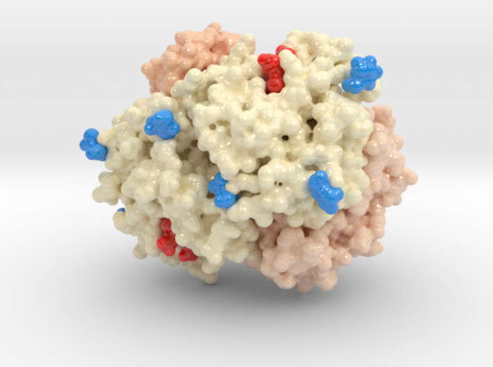
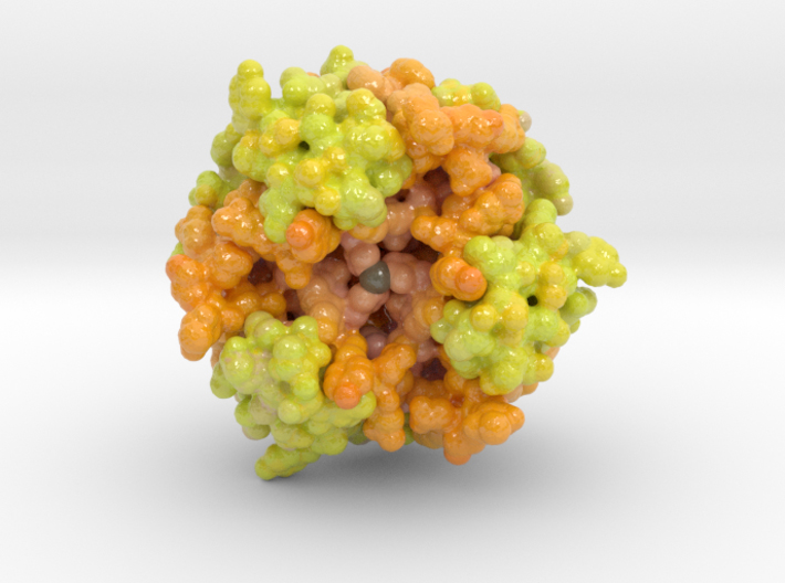
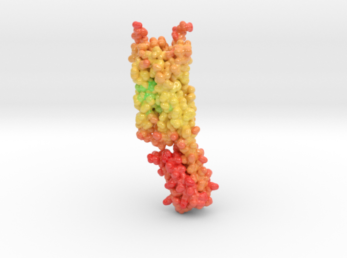
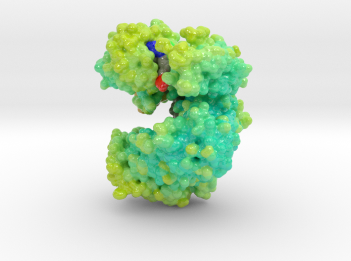
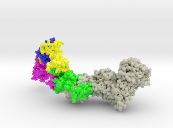
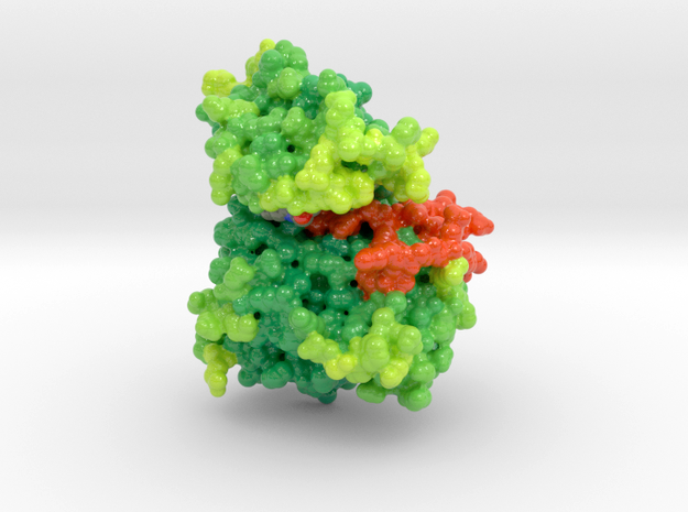
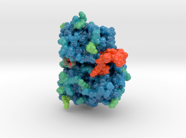
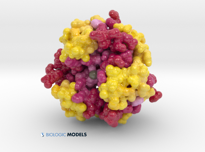
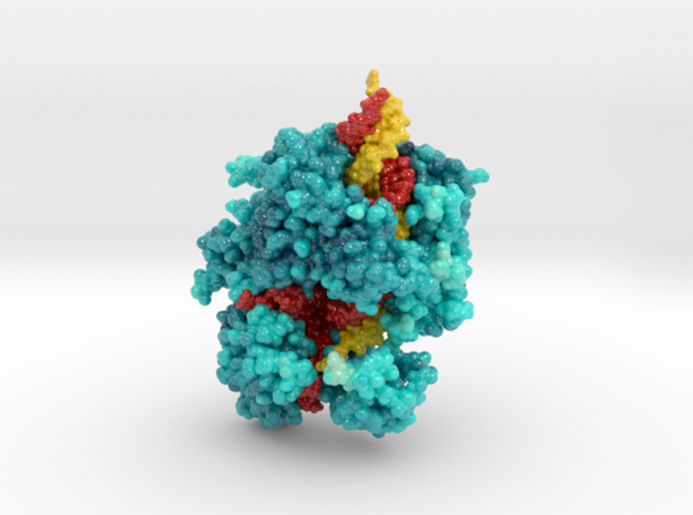
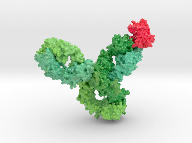
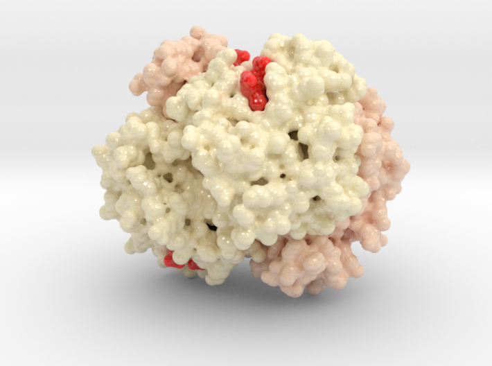
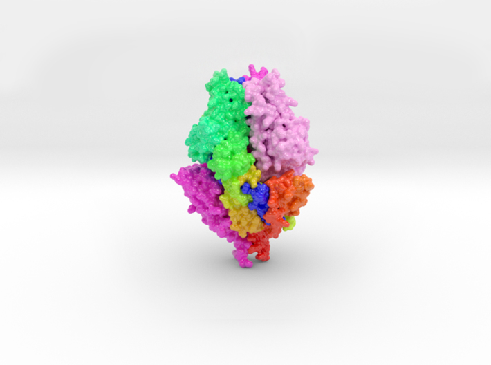
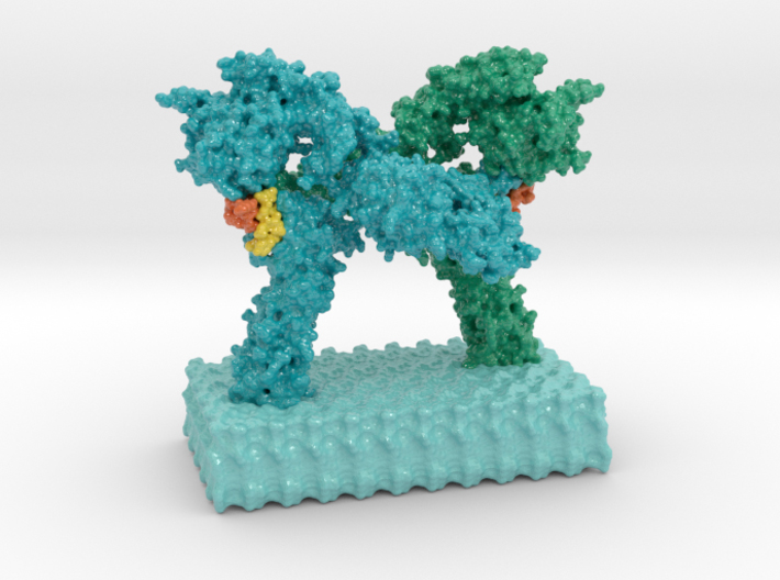
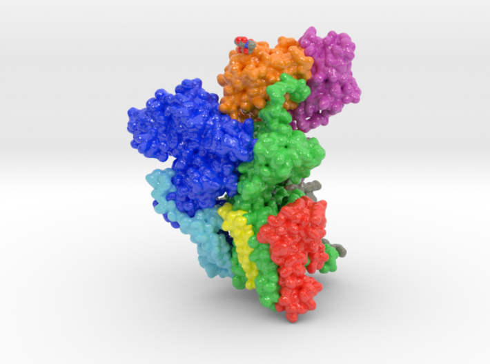
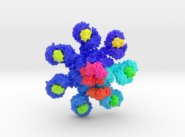
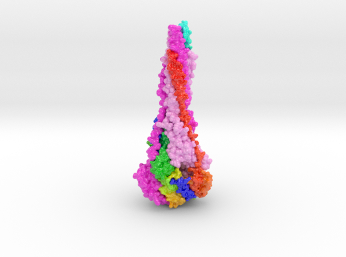
Deadly to newborn babies, the Human Respiratory Syncytial Virus (HSV) is a major cause of lower respiratory tract infection. Dynamic structural changes in the Fusion F Glycoprotein reveal and hide binding sites, making targets of new therapies difficult to identify. Explore RSV Fusion F Glycoprotein-3RRR and learn more about its Postfusion 3D structure. (more…)
Category: Volumetric
Biologic Models visualized by their ‘Volumetric’ surface.
Glycated Hemoglobin HbA1c
Human Insulin Hexamer
Psilocybin bound to Serotonin Receptor
Psilocybin bound to Serotonin Receptor Psilocybin, the active ingredient in psychoactive mushrooms, has provided the spiritual and cultural bedrock of many great civilizations. The Aztecs referred to teonanácatl, which translates as ‘divine mushroom’, and modern neuroscience has revealed how psilocybin interacts with serotonin receptors in the brain in order to produce a range of consciousness-altering…
Argonaute Protein
Argonaute Protein Small RNA sequences guide Argonaute Protein (Apo) to its specific DNA targets, which then leads to mRNA cleavage or translation inhibition. Inside the gut, bacteria express control over their host organism by secreting Nitric Oxide which modulates Apo function. Explore the 3D structure of Argonaute Protein. (more…)
Human Cytomegalovirus
The Human Cytomegalovirus (HCMV) is the leading cause of viral birth defects. HCMV Pentamer Complex is considered a key target candidate for vaccine therapies. How HCMV avoids humoral immunity is not clear. New research into the elusive virus reveals the surprising role of the host cell’s endoplasmic reticulum to aid viral infection. (more…)
Nucleosome 1KX5
TDP-43 Wild Type and Mutant in Complex with DNA
TDP-43 Wild Type and Mutant in Complex with DNA ALS is a fatal motor neuron disorder characterized pathologically by TAR DNA binding protein ( TDP-43 ). The function of TDP-43 is uncertain, and role in ALS remains speculative. Genome scanning reveals mutations associated to neural apoptosis. (more…)
Zika Virus Shell
EGFR Mutant Tyrosine Kinase
EGFR Mutant Tyrosine Kinase Mutations in EGFR lead to over-expression of proteins and different types of cancers as cells continually and proliferate. This 3D print of EGFR Tyrosine Kinase visualizes the mutations of key residues that lock the protein’s A-loop (orange) in its active conformation resulting in continued cellular proliferation. (more…)
EGFR Wild Type Tyrosine Kinase
Humalog Insulin Hexamer
CRISPR-Cas13a
Cytochrome MtrC
Monoclonal IgG4 Antibody bound to PD1
Monoclonal IgG4 Antibody bound to PD1 Humanized antibody are used in cancer immunotherapy to target the programmed cell death receptor (PD-1). Without the PD-1 protective mechanism, the antibody allows the immune system to destroy cancer cells. Explore the 3D structure with our interactive 3D models. (more…)
Oxygenated Hemoglobin Hb
Oxygenated Hemoglobin Hb Hemoglobin, the protein from inside red blood cells, transports oxygen molecules throughout the body. Its macro-molecular symmetrical structure just the first interesting characteristic of this fascinating molecule. Explore its 3D structure with our 3D printed protein models of Oxygenated Hemoglobin Hb. (more…)
RSV Prefusion Glycoprotein
This is a 3D print of the RSV Fusion Glycoprotein in its prefusion state. The 3D printed protein models of RSV Fusion F Glycoprotein visualize the conformational changes of this viral protein during membrane fusion. Two chains are colored shades of magenta, with the third chain colored blue to red from N to C-Terminal. (more…)
Insulin Receptor Complex
Oligosaccharyltransferase Complex
Oligosaccharyltransferase Complex This is a 3D print of oligosaccharyltransferase (OST) complex. It is colored blue according to the protein’s electrostatic property. Colored green are various monosaccharides attached to OST as sugar trees. Isolated and colored by atom type are the binding residues that attach shuttling proteins. Colored white are individual phospholipids from a plasma membrane.…