This is a 3D print of the RSV Fusion Glycoprotein in its prefusion state. The 3D printed protein models of RSV Fusion F Glycoprotein visualize the conformational changes of this viral protein during membrane fusion. Two chains are colored shades of magenta, with the third chain colored blue to red from N to C-Terminal.
Protein Description 5TDL
Bovine respiratory syncytial virus, a major cause of respiratory disease in calves, is closely related to human RSV, a leading cause of respiratory disease in infants. Recently, promising human RSV-vaccine candidates have been engineered that stabilize the metastable fusion (F) glycoprotein in its prefusion state; however, the absence of a relevant animal model for human RSV has complicated assessment of these vaccine candidates.
RSV Fusion Glycoprotein Realtime-3D Biologic Explorer
Use the interactive realtime 3D Biologic Explorer to visualize the internal anatomy and dynamic shape changes of the RSV Fusion Glycoprotein. (Best viewed in a full-screen desktop browser).
Model Description
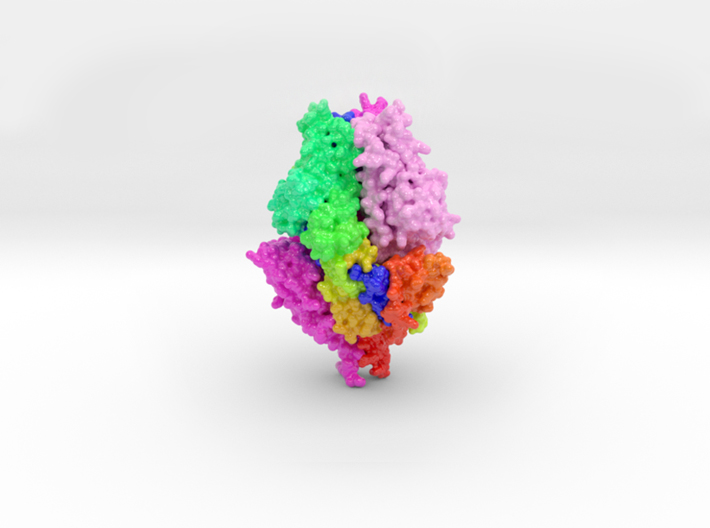
3D printed protein models of RSV Fusion F Glycoprotein visualize the conformational changes of this viral protein during membrane fusion. Each chain is colored uniquely, with one chain colored blue to red from N to C-Terminal.
3D Print RSV Fusion F Glycoprotein-5TDL
Using the interface below, configure the scale and materials using the horizontal slider and drop-down menu.