Posted on

Oxygenated Hemoglobin Hb

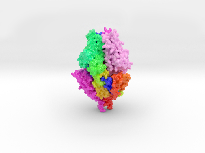
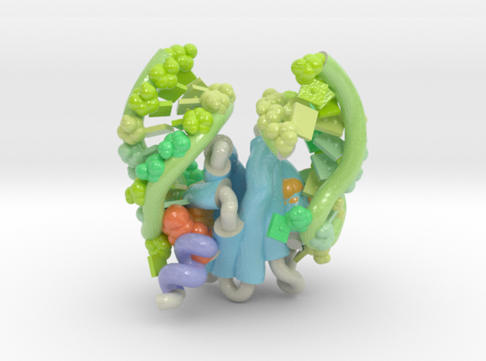
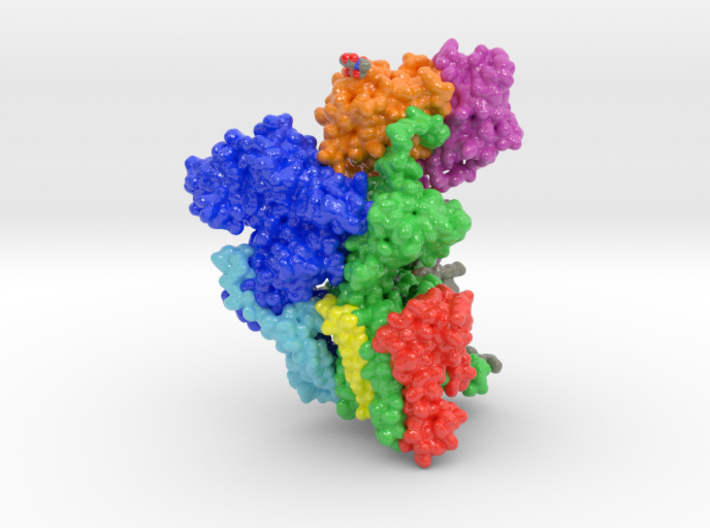
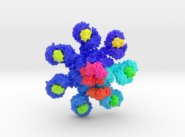
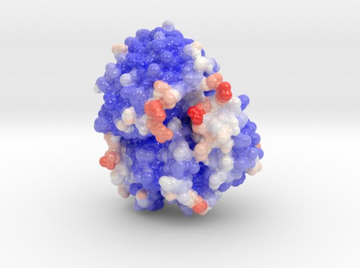
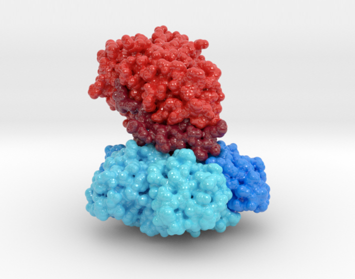
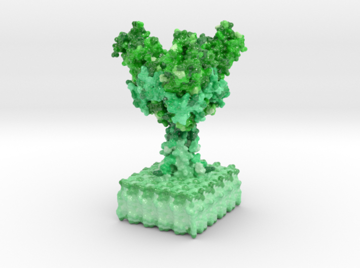
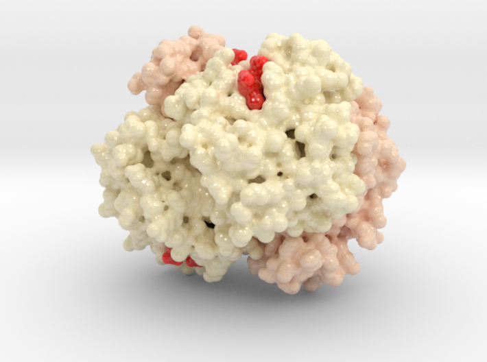
Oxygenated Hemoglobin Hb Hemoglobin, the protein from inside red blood cells, transports oxygen molecules throughout the body. Its macro-molecular symmetrical structure just the first interesting characteristic of this fascinating molecule. Explore its 3D structure with our 3D printed protein models of Oxygenated Hemoglobin Hb. (more…)

Read more

Posted on

Muscarinic Acetylcholine Receptor

Muscarinic Acetylcholine Receptor, 3UON

Muscarinic Acetylcholine Receptor 3UON Muscarinic acetylcholine receptors are G protein-coupled receptor complexes in the cell membranes of certain neurons and cells. They play several roles, including acting as the main end-receptor stimulated by acetylcholine released from postganglionic fibers in the parasympathetic nervous system. This 3D printed protein model of muscarinic acetylcholine receptor is visualized by its ribbon structure highlighting the protein’s transmembrane active sites. (more…)

Read more

Posted on

RSV Prefusion Glycoprotein

RSV, Fusion, Glycoprotein, Prefusion, 5TDL

This is a 3D print of the RSV Fusion Glycoprotein in its prefusion state. The 3D printed protein models of RSV Fusion F Glycoprotein visualize the conformational changes of this viral protein during membrane fusion. Two chains are colored shades of magenta, with the third chain colored blue to red from N to C-Terminal. (more…)

Read more

Posted on

FAB Antibody bound to Meningococcal fHbp

fHbp, Fusion H-binding protein, Antibody, FAB, Biologic Models

FAB Antibody bound to Meningococcal fHbp 3D printed protein models of a FAB Antibody bound to Meningococcal fHbp. These models are designed to pair with the academic publication, Crystal structure reveals vaccine elicited bactericidal human antibody targeting a conserved epitope on meningococcal fHbp. (LINK ) (more…)

Read more

Posted on

Oligosaccharyltransferase Complex

Oligosaccharyltransferase Complex, OST, ETH Zurich, Biologic Models

Oligosaccharyltransferase Complex This is a 3D print of oligosaccharyltransferase (OST) complex. It is colored blue according to the protein’s electrostatic property. Colored green are various monosaccharides attached to OST as sugar trees. Isolated and colored by atom type are the binding residues that attach shuttling proteins. Colored white are individual phospholipids from a plasma membrane.…

Read more

Posted on

Ebola Virus Ectodomain

Ebola, Virus, Biologic Models

Ebolavirus (EBOV) is a highly virulent pathogen capable of causing a severe hemorrhagic fever with 50–90% lethality. The EBOV glycoprotein (GP) is the only virally expressed protein on the virion surface and is critical for attachment to host cells. 3D print model the ectodomain of the Ebolavirus. (more…)

Read more