Poliovirus Type 2
This is 3D protein model of the Poliovirus Type 2 3D printed in full-color sandstone or plastic and colored to denote the locations of active residues targeted by antibodies. Despite the Type 2 wild poliovirus being declared eradicated in September 2015, a vaccine-derived mutated version of the vaccine-virus has reemerged in multiple countries.
Virus Description
Polioviruses are human pathogens that affects the central nervous system and can cause temporary or permanent paralysis. Polioviruses are icosahedral single-stranded RNA viruses, which belong to the picornavirus family, and occur as three distinct serotypes. All three serotypes of poliovirus can infect primates, but only type 2 can infect mice. — NCBI Citation
Biologic Explorer: Poliovirus Type 2 Shell 1EAH
Explore x-ray crystallographt dataset 1EAH and examine the underlying protein structure.
Http iframes are not shown in https pages in many major browsers. Please read this post for details.Polio Disease and the Poliovirus
Polio disease is caused by viral infection that eventually leads to paralysis throughout the body. There are three wild types of poliovirus (WPV) – type 1, type 2, and type 3. People need to be protected against all three types of the virus in order to prevent polio disease and the polio vaccination is the best protection. The Global Polio Laboratory Network (GPLN) has confirmed the isolation of type 2 vaccine-derived poliovirus (VDPV2) from environmental samples collected from multiple countries around the world.
Molecular Visualization
How did the Vaccine-Derived Poliovirus Emerge
There are two types of poliovaccines, live-attenuated virus vaccines and inactived virus vaccines. For many years, the oral polio vaccine (OPV) that has brought the wild poliovirus to the brink of eradication. The oral polio vaccine is based on a live attenuated (weakened) version of the virus. This type of vaccine provides better immunity in the gut, which is where polio replicates. The vaccine-virus is also excreted in the stool, and in communities with low-quality sanitation, this means that even though the vaccine-virus is weakened, it can be spread from person to person. While this can actually help protect the community, if this virus mutates and becomes more virulent, it can regain hold within communities with low immunization rates. This mutated form that can cause paralysis just like the wild poliovirus.
Effective strategies including inhibitory antibodies and small molecules are currently under design to target VDPV2 virions.
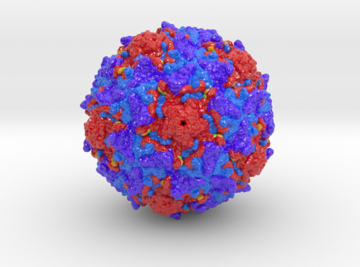
Biologic Explorer: Poliovirus Type 2 Capsid 1EAH
The primary capsid subunit of the virus that is repeated sequentially around the virus core is composed of 4 viral proteins (VP1: red, VP2: purple, VP3: blue, and VP4: orange). Understanding the structural dynamics of the poliovirus during receptor binding is key to developing therapeutic antibodies that block viral function. Viral Protein 1 (red) offers primary polar charged binding sites K214 and E224 (colored green and yellow) that bind the CD155 receptor and therefore function as targets for therapeutic intervention.
Http iframes are not shown in https pages in many major browsers. Please read this post for details.3D Print Poliovirus Type 2 Shell
Purchase original 3D prints of the Poliovirus in either Full-color Sandstone or high definition Full-color Plastic.