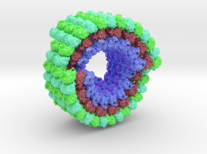
3D Print of Microtubule 3J2I custom colored to visualize the Tubulin 1A-2B complex (Blue) at the core of the microtubule and another Tubulin 1A-2B complex wraps around the outer shell (Green) and separated by Kinesin (red) molecules.
To elucidate the structural basis of the mechanism of microtubule depolymerization by kinesin-13s, we analyzed complexes of tubulin and the Drosophila melanogaster kinesin-13 KLP10A by electron microscopy (EM) and fluorescence polarization microscopy. We report a nanometer-resolution (1.1 nm) cryo-EM three-dimensional structure of the KLP10A head domain (KLP10AHD) bound to curved tubulin. We found that binding of KLP10AHD induces a distinct tubulin configuration with displacement (shear) between tubulin subunits in addition to curvature. In this configuration, the kinesin-binding site differs from that in straight tubulin, providing an explanation for the distinct interaction modes of kinesin-13s with the microtubule lattice or its ends. The KLP10AHD-tubulin interface comprises three areas of interaction, suggesting a crossbow-type tubulin-bending mechanism. These areas include the kinesin-13 family conserved KVD residues, and as predicted from the crossbow model, mutating these residues changes the orientation and mobility of KLP10AHDs interacting with the microtubule.
Biologic Explorer: 3J2U
To better understand the structural relationship of domain within the Spike Glycoprotein, a protein dataset 7KS9 has been colorized to match our 3d printed model. Explore each domain below after “Initializing” the dataset.
Http iframes are not shown in https pages in many major browsers. Please read this post for details.3D Print Microtubule 3J2U
Biologic Model of Microtubule 3J2U 3D printed in Full-color Sandstone, Nylon, and Plastic. 3D Print of Microtubule 3J2I custom colored to visualize the Tubulin 1A-2B complex (Blue) at the core of the microtubule and another Tubulin 1A-2B complex wraps around the outer shell (Green) and separated by Kinesin (red) molecules. Tryptophane residues interconnect the Tubulin molecules and residues from each chain tinted darker shade of the chain color.
Custom 3D Printed Protein Model
Interested in 3D printing a different molecule? Submit a 3D print request and one of our visualization scientists will help you create it. [otw_shortcode_button href=”https:///biologicmodels.com/3D-print-request” size=”medium” icon_position=”left” shape=”square”]Submit Request[/otw_shortcode_button]