Glycated Hemoglobin HbA1c
Learn more about the dangers of high blood sugar and the HbA1c blood test by exploring this 3D Printed Protein Model of Glycated Hemoglobin (HbA1c).
Protein Description 3B75
Glycated Hemoglobin (HbA1c) is a derivative compound that results when Hemoglobin (Hb) is exposed to high levels of blood sugar for prolonged periods of time. Sugar is extremely cytotoxic and readily binds to proteins throughout the body. Glycosylation causes structural changes in the protein, preventing Hemoglobin from achieving optimal 3D conformation and biochemical function.
Protein Location
Hemoglobin the most abundant protein inside red blood cells and transporter oxygen molecules throughout the body. Red blood cells aren’t the only location we find Hemoglobin in nature. Various forms of Hemoglobin can be found throughout the body performing various functions other than transporting oxygen. Hemoglobin can even be found in plants, and even legumes. In plants, oxygen molecules can damage the molecular function of some proteins. Hemoglobin works to protect these molecular machines by savaging for oxygen, preventing damage.

3D Print Glycated Hemoglobin HbA1c 3B75
Model Description
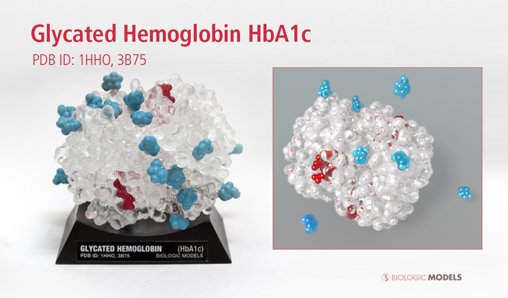
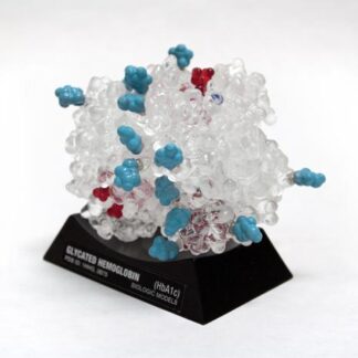
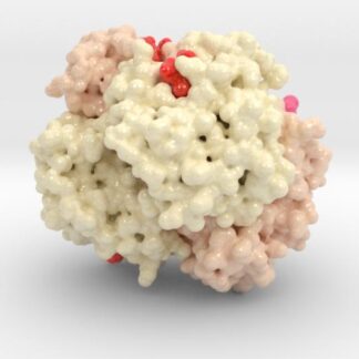
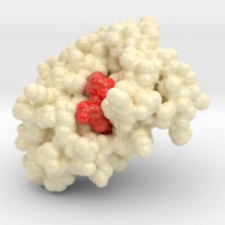
This 3D printed protein model of Oxygenated Hemoglobin Hb 1HHO color-coded to show each chain of the protein (Shades of pink) and the 4 HEME Groups (Red) that bind Oxygen Molecules (dark blue unseen). Glycated Hemoglobin HbA1c 3B75 showcases the effects of prolonged exposure to elevated blood glucose (Cyan). Model created from PDB ID: 1HHO, 3B75.
Protein Structure
Hemoglobin is a relatively large protein, it’s reported resolution is 2.1 Å. Composed of four protein chains, 2 Alpha chains and 2 Beta chains, each chain has a single ring-like HEME Group (red x 4) with a single iron atom in its core. Attached across its surface are glucose and fructose molecules that bind to multiple residues across the protein’s surface.
Biologic Explorer: 3B75
Explore the protein dataset used to create the 3D printed protein model of Glycated Hemoglobin HbA1c 3B75.
Hemoglobin Anatomy
| Structure | Residues | Atoms |
| Chain α | 141 | 1069 (x 2) |
| Chain ß | 146 | 1123 (x 2) |
| HEME Group | 1 | 43 (x 4) |
| Oxygen | 1 | 2 (x 4) |
| Glucose | 1 | 12 (x 16) |
Glycated Hemoglobin A1c
Hemoglobin makes for an ideal target to determine long-term exposure to elevated levels of glucose, because Hemoglobin is easily drawn from the body in blood samples. When the blood stream is saturated with too much sugar, hemoglobin begins to accumulate sugar particles on its surface. Hemoglobin is a strong and robust protein, but even it suffers marginally because of sugar accumulation. To our scientific advantage, the accumulation of sugar on hemoglobin can be measured. From this measurement, we gain insights
The Hemoglobin A1c Diabetes Blood Test
When sugar binds to hemoglobin, it’s a permanent bond that doesn’t break. This means that as long as that particle of Hemoglobin is still circulating in that blood cell, hemoglobin collects more sugar. Because red blood cell’s lifespan is only about 3 months, the Glycated Hemoglobin A1c test is a great snapshot of blood glucose control for the last 3 months.
By simplifying a complex biochemical process, the material interactions of the model relayed intuitively the dangers of high blood sugar, while the educator relayed positive self-management practices.
The Glycated Hemoglobin HbA1c blood test, or more commonly known as the “A1c” blood test, is an important test given to people around the world, many of those at risk of developing diabetes. The test is the universally preferred method of diagnosing the onset of type 2 pre-diabetes and the only blood test that does not need overnight fasting. Achieving improved A1C scores is an important milestone in lifestyle intervention programs focused on reversing pre-diabetes.
Photos of the Interactive Glycated Hemoglobin HbA1c
As important as this blood test is, the A1C blood test is universally misunderstood; too often confused with acute blood sugar readings. The Interactive Hemoglobin A1C protein model solves this problem by transforming a misunderstood blood test result into an interactive scientific experience. The interactive model simulates the glycosylation of hemoglobin using magnets to bind glucose (blue) directly to the model surface.
Explaining the A1c Test to Kids Living with Type 1 Diabetes
Shortly after completing the design and fabrication of the Hemoglobin A1c Teaching Model, we tested the effectiveness of using it to teach children living with type 1 and 2 diabetes about the HbA1c test. The goal of the experiment was to explain to children the importance of checking their blood glucose regularly, explain what the HbA1c blood test analyses, and how prolonged exposure to glucose causes high HbA1c scores. We contacted the Diabetes Youth Foundation of Indiana to see if they’d like to try using our model during their yearly summer camp retreat.
HbA1c Model questionnaire taken by children at diabetes summer camp
Four age groups of children were introduced to the model during their a lecture explaining the HbA1c test. The children’s diabetes educator used the magnetized pieces of glucose (blue) to show how sugar binds to proteins, letting the magnetic properties of the materials explain glycosylation. To represent increased HbA1c scores, the educator would attach more glucose pieces to the model. By simplifying a complex biochemical process, the material interactions of the model relayed intuitively the dangers of high blood sugar, while the educator relayed positive self-management practices. After the class, the children were asked to answer a few questions about the A1c test and the information they learned while playing with the model. The results were more than fantastic. The diabetes educator reported that every single child took away new information about the test and the importance of good blood glucose control.
Model Descriptions
Biologic Models has a variety of Hemoglobin based models. In specific, we have two types of Glycated Hemoglobin A1c and Oxygenated Hemoglobin, each in different materials and sizes. Please see the description below to learn more about each product and functionality.
3D Print Hemoglobin Models
Models available in both Full-color Sandstone and Full-color Plastic.
Interactive Glycated Hemoglobin A1c Model (Acrylic)
This model of Hemoglobin A1c is based on x-ray crystallography data sets: 1HHO, and 3B75. These models are cast by hand in high quality, durable and washable plastics. Model dimensions are 4in x 3cm x 3in. Each model comes with a custom made display base.
3D Print Other Hemoglobin Models
This is a 3D print of Glycated Hemoglobin (HbA1c), created from PDB ID: 3B75. The globulin chains (2x A,B) of this model are colored white, HEME groups red, and Oxygen Molecules Blue. This model also visualizes the HbA1c diabetes blood test by showing how sugar molecules (cyan) attach to proteins changing their shape and decrease its functionality. Based on simulations of potential glycation sites, we have isolated the Lys binding residues (medium Grey) and attached sugar molecules. When compared to our normal Oxygenated Hemoglobin (Hb), you can quickly explain both what the HbA1c blood test examines, as well as why it is so important for patients to control their blood glucose.
Custom 3D Print Request
Request a custom 3D printed protein model. Send us the protein name and PDB ID of the protein you’re interested in printing and we’ll get back to you with a feasibility analysis and estimate for printing.
[ninja_form id=6]


