EGFR Wild Type Tyrosine Kinase
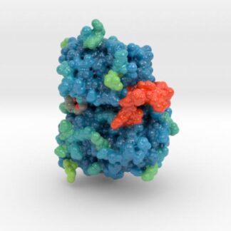
This is a 3D print of X-ray crystallography dataset 4HJO, the Crystal Structure of the Inactive EGFR Tyrosine Kinase Domain with Erlotinib.
Protein Description
Epidermal growth factor receptor (EGFR) exists on the cell surface and is activated by binding of its specific ligands, including epidermal growth factor and transforming growth factor α (TGFα) (note, a full list of the ligands able to activate EGFR and other members of the ErbB family is given in the ErbB article). ErbB2 has no known direct activating ligand, and may be in an activated state constitutively or become active upon heterodimerization with other family members such as EGFR. Upon activation by its growth factor ligands, EGFR undergoes a transition from an inactive monomeric form to an active homodimer.
Biologic Explorer: 4HJO
Http iframes are not shown in https pages in many major browsers. Please read this post for details.
Dataset Description
“Erlotinib and gefitinib, tyrosine kinase inhibitors used to block EGFR (epidermal growth factor receptor) signaling in cancer, are thought to bind only the active conformation of the EGFR-TKD (tyrosine kinase domain). Through parallel computational and crystallographic studies, we show in the present study that erlotinib also binds the inactive EGFR-TKD conformation, which may have significant implications for its use in EGFR-mutated cancers.”
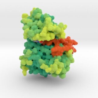
Inactive EGFR Tyrosine Kinase 3D Molecular Visualization
This 3D animation depicts EGFR bound by Erlotinib. Important protein structures like the alphaC-Helix, P-Loop and A-Loop are visualized to identify EGFR’s active state.
Inactive EGFR 3D Visualization

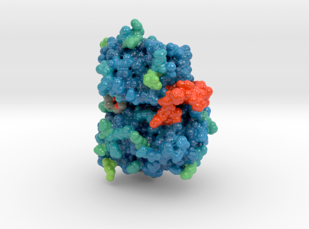
3D Print of Inactive EGFR Tyrosine Kinase
If you’d like to explore EGFR, you can purchase a 3D print of EGFR in both its active and inactive state. Models available in multiple sizes and materials.