Oxygenated Hemoglobin Hb
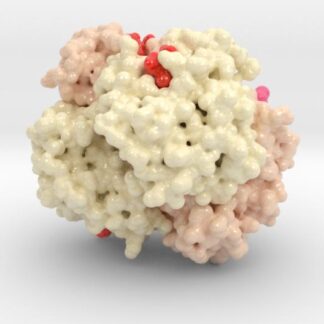
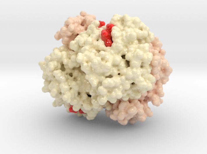
Hemoglobin, the protein from inside red blood cells, transports oxygen molecules throughout the body. Its macro-molecular symmetrical structure just the first interesting characteristic of this fascinating molecule. Explore its 3D structure with our 3D printed protein models of Oxygenated Hemoglobin Hb.
Protein Description 1HHO
Hemoglobin; abbreviated Hb or Hgb, is the iron-containing oxygen-transport metalloprotein in the red blood cells of all vertebrates as well as the tissues of some invertebrates. Hemoglobin in the blood carries oxygen from the lungs or gills to the rest of the body (i.e. the tissues). There it releases the oxygen to permit aerobic respiration to provide energy to power the functions of the organism in the process called metabolism. A healthy individual has “12 to 16” grams of hemoglobin in every 100 ml of blood.
In 1825 J. F. Engelhard discovered that the ratio of iron to protein is identical in the hemoglobins of several species. This protein model was created from PDB ID: 1HHO
Biologic Explorer: 1HHO
Http iframes are not shown in https pages in many major browsers. Please read this post for details.
Model Descriptions
Biologic Models has a variety of Hemoglobin based models. In specific, we have two types 3D printed models of Glycated Hemoglobin A1c and Oxygenated Hemoglobin models, each in different materials and sizes. As well, we have High-quality acrylic models of Glycated Hemoglobin and Oxygenated Hemoglobin. Please see the descriptions below to learn more about each product and functionality.
3D Print Hemoglobin Models
Models available in both Full-color Sandstone and Full-color Plastic.
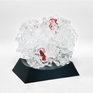
Interactive Glycated Hemoglobin A1c Model (Acrylic)

This model of Hemoglobin A1c is based on x-ray crystallography data sets: 1HHO, and 3B75. These models are cast by hand in high quality, durable and washable plastics. Model dimensions are 4in x 3cm x 3in. Each model comes with a custom made display base.
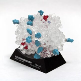
3D Print Other Hemoglobin Models
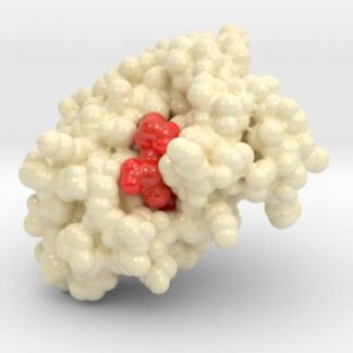
This is a 3D print of Glycated Hemoglobin (HbA1c), created from PDB ID: 3B75. The globulin chains (2x A,B) of this model are colored white, HEME groups red, and Oxygen Molecules Blue. This model also visualizes the HbA1c diabetes blood test by showing how sugar molecules (cyan) attach to proteins changing their shape and decrease its functionality. Based on simulations of potential glycation sites, we have isolated the Lys binding residues (medium Grey) and attached sugar molecules. When compared to our normal Oxygenated Hemoglobin (Hb), you can quickly explain both what the HbA1c blood test examines, as well as why it is so important for patients to control their blood glucose.