EGFR Tyrosine Kinase
An investigation of the structural differences between wild type and mutant EGFR Tyrosine Kinase in order to understand oncogenic cellular proliferation.
Protein Description
EGFR is an important protein for a variety of different types of oncology. It is important for the development of mammary glands. It’s ligands ( TGF-α, and heregulin) play such vital roles in development, that in the absence of hormone signals can still induce ductal and lobuloalveolar development.
Protein Location


EGFR is located on the surface of cells throughout the body. When activated by different cytokines, receptors dimerize and initiates downstream signaling via its intracellular tyrosine kinase domains.
EGFR Visualization
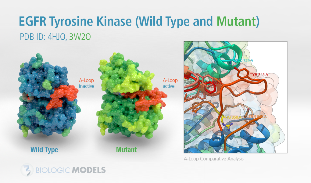
Protein Structure
Dimerization stimulates the intracellular tyrosine kinase domains. This begins a process where several tyrosine residues are autophosphorylated. (Y992, Y1068, Y1148, Y1173 ). Downstream signaling following this includes MAPK, Akt, and HNK pathways. These lead to both DNA synthesis and cellular proliferation. If this process becomes uncontrolled, an unfortunate disease state develops.
Inactive EGFR 3D Animation

3-Dimensional States
Tyrosine Kinases exist in two distinct states, inactive and active. Inactive states are marked by their contracted a-loop with a closed N-lobe. When closed, the ATP binding pocket of the tyrosine kinase is blocked. and tyrosine residues are protected from phosphorylation. In the active state, the a-loop is extended with an open N-lobe revealing the ATP-binding pocket of the kinase.
Photo Gallery


Role in Health and Disease
Cancer
Mutations in EGFR lead to overexpression of proteins and different types of cancers as cells continually and proliferate. Advancements in drug design allow for the continued binding of small molecules despite mutations. These compounds prevent kinase phosphorylation and down-regulate EGFR signals.
Mutant Active EGFR 3D Animation
Inflammatory Disease
Cancer isn’t the only issue. Psoriasis, eczema, and atherosclerosis are all implicated in over-expression of EGFR. This is not well understood yet, but likely due to over-expression of inflammatory cytokines that initiate EGFR phosphorylation.
Drug Therapies
A common strategy of small molecule inhibitors is to identify receptors and examine their intra-cellular kinase domains. In many cases, this prevents aberrant behaviors in the receptor and mitigate the develop of disease. An easy target for this inhibition process is the kinase domain’s ATP binding pocket. It has a high affinity for its ligand and phosphorylation of its tyrosine residue a requirement for signal propagation.
3D Print EGFR Tyrosine Kinase
3D printed protein models available in multiple sizes and materials. Contact us if you’d like to request a customization: CONTACT