Description
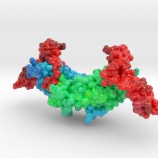
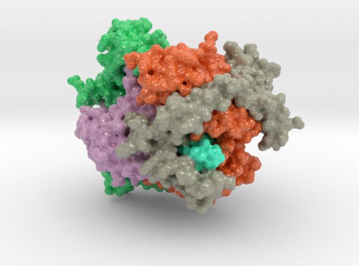
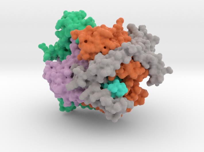
The structure of omphalotin A (OphA) reveals a complex catenane-like arrangement in which the peptide substrate is clamped with its amide nitrogen aligned for nucleophilic attack on the methyl group of SAM.
Model Description
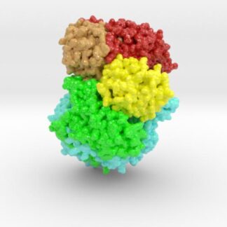
This 3D printed protein model of Omphalotin A (OphA) Dimer. This protein model set was designed to pair with the publication listed below matching Figure.1
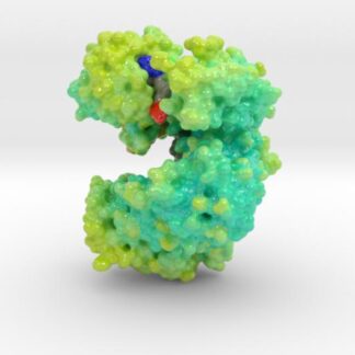
The dimer has a pseudo interlocking ring (catenane) arrangement. The first monomer of the two chain protein structure is colored to visualize the methyltransferase domain (orange), the clasp domain (green), and C-terminal substrate peptide (cyan). The active site of OphA and the ligand S-Adenosylmethionne (SAM) are colored by their atom type. (carbon = orange/yellow, nitrogen = blue and oxygen = red.)
The second chain of the dimer colors the same regions in different colors. The other monomer is colored pink (N-terminal methyltransferase domain) and gray (C-terminal clasp domain). The clasp terminal domain wraps around the loops that cover the active site of the other monomer.
For international customers outside the US, please visit the model on our 3D printing service’s international website: LINK
Select the desired material finish and size below. Matte finish applies a UV protective, semi-glossy coating. Natural finish does not.
Created from PDB ID: 5N0S